
Oral Diagnosis: Minimally Invasive Imaging Approaches for Accurate Diagnosis

In the field of dentistry, accurate diagnosis is crucial for developing effective treatment plans and ensuring optimal patient outcomes. Traditional imaging techniques, such as panoramic radiography and intraoral radiography, have been widely used for decades. However, these techniques often involve exposure to ionizing radiation, which can pose potential health risks. Additionally, they may not provide sufficient detail for complex dental conditions.
To overcome these limitations, minimally invasive imaging approaches have emerged as valuable tools in oral diagnosis. These techniques offer several advantages, including reduced radiation exposure, enhanced image quality, and the ability to visualize anatomical structures in three dimensions. This article provides a comprehensive overview of minimally invasive imaging techniques used in oral diagnosis, discussing their applications, advantages, limitations, and impact on treatment planning.
4.4 out of 5
| Language | : | English |
| File size | : | 22899 KB |
| Text-to-Speech | : | Enabled |
| Screen Reader | : | Supported |
| Enhanced typesetting | : | Enabled |
| Print length | : | 234 pages |
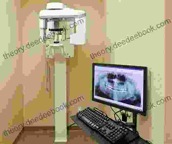
Cone Beam Computed Tomography (CBCT)
Cone beam computed tomography (CBCT) is a three-dimensional (3D) imaging technique that uses a cone-shaped beam of X-rays to capture cross-sectional images of the jaws and surrounding structures. CBCT provides detailed and accurate images of dental anatomy, including teeth, bone, and soft tissues, making it a valuable tool for diagnosing and treatment planning a wide range of dental conditions.
Advantages of CBCT:
- High-resolution images: CBCT produces high-resolution images that allow dentists to visualize anatomical structures in great detail.
- 3D reconstruction: CBCT images can be reconstructed into 3D models, providing a comprehensive view of the jaws and surrounding tissues.
- Reduced radiation exposure: CBCT uses a lower dose of radiation compared to traditional CT scans, minimizing the potential health risks associated with ionizing radiation.
- Wide range of applications: CBCT is used in a variety of dental applications, including implant planning, root canal treatment, and orthodontic diagnosis.
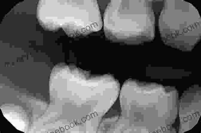
Dental Radiography
Dental radiography, also known as X-rays, is a widely used imaging technique that produces two-dimensional (2D) images of the teeth and surrounding structures. Dental X-rays are commonly used to detect cavities, periodontal disease, and other dental abnormalities.
Types of Dental Radiography:
- Bitewing radiographs: Bitewing X-rays are used to visualize the crowns of the teeth and detect cavities between the teeth.
- Periapical radiographs: Periapical X-rays are used to visualize the entire tooth, including the root and surrounding bone.
- Panoramic radiographs: Panoramic X-rays provide a wide view of the entire jaw, including the teeth, bone, and surrounding structures.
Advantages of Dental Radiography:
- Widely available: Dental X-rays are widely available in dental offices and clinics.
- Low cost: Dental X-rays are relatively inexpensive compared to other imaging techniques.
- Fast and convenient: Dental X-rays can be taken quickly and easily.
- Useful for detecting cavities and periodontal disease: Dental X-rays are effective in detecting cavities between the teeth and periodontal bone loss.
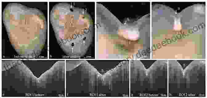
Optical Coherence Tomography (OCT)
Optical coherence tomography (OCT) is a non-invasive imaging technique that uses light waves to create cross-sectional images of biological tissues. OCT provides high-resolution images of the microstructure of tissues, allowing dentists to visualize the layers of the tooth and detect early signs of decay and other dental conditions.
Advantages of OCT:
- Non-invasive: OCT does not involve ionizing radiation, making it a safe and comfortable imaging technique.
- High-resolution images: OCT produces high-resolution images that allow dentists to visualize the microstructure of tissues in great detail.
- Early detection of decay: OCT can detect early signs of tooth decay before it becomes visible on X-rays.
- Monitoring treatment: OCT can be used to monitor the progression of dental treatments, such as root canal therapy.
Impact on Treatment Planning
Minimally invasive imaging approaches have revolutionized treatment planning in dentistry. These techniques provide dentists with detailed and accurate information about the patient's anatomy, allowing them to make informed decisions and develop optimal treatment plans. For example:
CBCT:
CBCT is essential for planning dental implants. The 3D images provided by CBCT allow dentists to assess the bone density and volume, determine the ideal implant size and placement, and avoid potential complications.
Dental Radiography:
Dental X-rays are used to detect cavities, periodontal disease, and other dental abnormalities. This information is crucial for developing treatment plans that address the specific needs of the patient.
OCT:
OCT can be used to monitor the progression of dental treatments, such as root canal therapy. By visualizing the microstructure of the tooth, dentists can assess the effectiveness of the treatment and make necessary adjustments to ensure a successful outcome.
Minimally invasive imaging approaches have become indispensable tools in oral diagnosis. These techniques offer reduced radiation exposure, enhanced image quality, and the ability to visualize anatomical structures in three dimensions. By providing dentists with detailed and accurate information, minimally invasive imaging approaches enable precise diagnosis, optimal treatment planning, and improved patient outcomes.
4.4 out of 5
| Language | : | English |
| File size | : | 22899 KB |
| Text-to-Speech | : | Enabled |
| Screen Reader | : | Supported |
| Enhanced typesetting | : | Enabled |
| Print length | : | 234 pages |
Do you want to contribute by writing guest posts on this blog?
Please contact us and send us a resume of previous articles that you have written.
 Novel
Novel Page
Page Chapter
 Text
Text Paperback
 E-book
 Magazine
 Sentence
 Bookmark
 Bibliography
 Synopsis
 Annotation
 Footnote
 Manuscript
 Codex
Codex Bestseller
 Library card
 Biography
 Autobiography
 Memoir
 Reference
 Dictionary
 Thesaurus
 Resolution
 Stacks
 Archives
 Periodicals
 Study
Study Lending
 Reserve
 Journals
 Special Collections
 Interlibrary
 Dissertation
 Storytelling
 Awards
 Reading List
 Book Club
 Theory
 Textbooks
 R W Swartz
 Leo Carew
 Paul Blakey
 Mark Bryant
 Peter Hough
 Kensuke Suzuki
 Richard Scase
 Zhihua Zhang
 George W Breslauer
 Demarius Jackson
 Tracy Brown
 Heather Robinson
 Marion Milton
 I P Mayers
 Fawn Weaver
 Malcolm Slesser
 Paul Larosa
 Charles Dagher
 Sarra Jenkins
 Dean J Kotlowski
Light bulbAdvertise smarter! Our strategic ad space ensures maximum exposure. Reserve your spot today!

 Jimmy ButlerBeginnings Beyond Foundations: Exploring Cutting-Edge Innovations in Early...


 Albert CamusFollow ·17k
 Isaias BlairFollow ·12k
 Don ColemanFollow ·13.3k
 Jerome BlairFollow ·13.2k
 Cole PowellFollow ·14.6k
 Jayden CoxFollow ·5.4k
 Jacques BellFollow ·16.3k
 Chad PriceFollow ·12.9k

 Charlie Scott
An Extensive Guide to Road Races in the Southern United...
Welcome to the...
 Seth Hayes
How to Create Your Cosmetic Brand in 7 Steps: A...
The cosmetic industry is booming, with an...
 Emilio Cox
Lean for Dummies: A Comprehensive Guide to the Lean...
Lean is a management...

 Dashawn Hayes
The Family She Never Met: An Enthralling Novel of...
Prologue: A Serendipitous...

 Italo Calvino
The Alluring Soundscape of Rickie Lee Jones: A Journey...
: The Enigmatic Soul of...

 Fyodor Dostoevsky
For The Love Of Dylan: An Exploration of Bob Dylan's...
Bob Dylan, the...
4.4 out of 5
| Language | : | English |
| File size | : | 22899 KB |
| Text-to-Speech | : | Enabled |
| Screen Reader | : | Supported |
| Enhanced typesetting | : | Enabled |
| Print length | : | 234 pages |
